
22 week 3D Ultrasound Pictures A Date With Baby
Contents. 1 What to Expect and Why the 26-Week 3D Ultrasound is an Important Milestone in Pregnancy. 1.1 What to Expect During a 26 Week 3D Ultrasound; 1.2 Understanding the Importance of a 26 Week 3D Ultrasound. 1.2.1 Early Detection of Potential Health Issues; 1.2.2 Bonding Experience for Expectant Parents; 1.2.3 Preparation for the Arrival of the Baby; 1.3 FAQ about topic 26 Week 3D.

28 Weeks Ultrasound
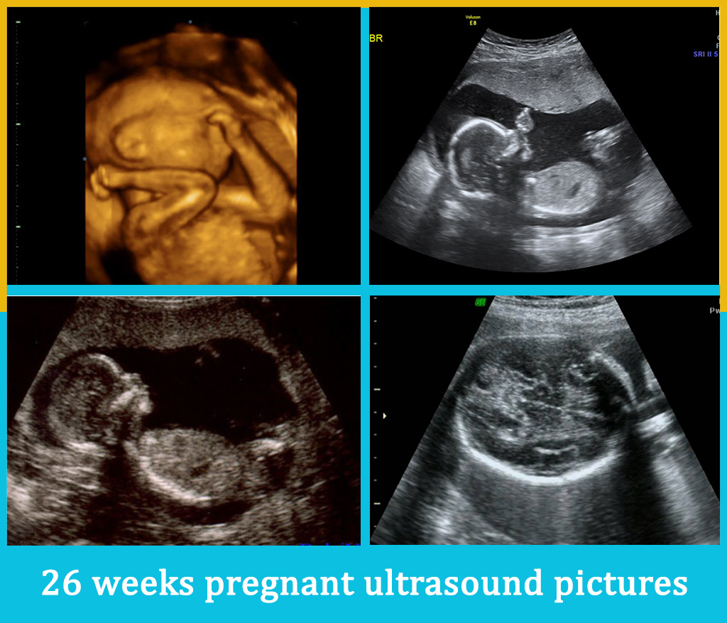
Your Baby's Development at 26 Weeks . At 26 weeks, a baby is almost 9 1/4 inches (23.4 centimeters) from the top of their head to the bottom of the buttocks (known as the crown-rump length), and baby's height is about 13 inches (33.3 centimeters) from the top of the head to the heel (crown-heel length). This week, baby weighs just about.
3D 4D 5D HD Ultrasound Packages Early Gender
OUR 3D/4D ULTRASOUND AT 26 WEEKS + HUSBAND SEES BABY FOR THE FIRST TIME!!! (UC BABY) Beth Grace Moore 43.5K subscribers Subscribe 5.4K views 2 years ago #secondtrimester #3dultrasound.
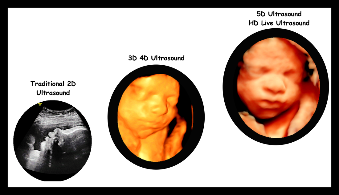
Still confused about the difference between 3D 4D and 5D ultrasound?
Providers use abdominal ultrasounds after about 12 weeks of pregnancy. Traditional ultrasounds are 2D. More advanced technologies like 3D or 4D ultrasound can create better images. This is helpful when your provider needs to see your baby's face or organs in greater detail. Not all providers have 3D or 4D ultrasound equipment or specialized.
3D/4D Ultrasound Packages & Pricing UltraBaby
Short answer 26 weeks ultrasound: A 26-week ultrasound is a routine prenatal screening test to check the baby's growth, development, and position in the womb. It also allows doctors to assess the placenta, amniotic fluid levels, and detect any abnormalities or potential complications. The procedure uses high-frequency sound waves to produce images of the fetus
Mommy in the Making Wordless Wednesday Ultrasound pics!
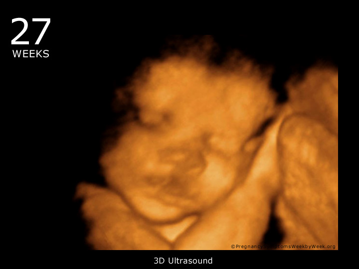
No, 26 weeks is actually an ideal time to schedule a 3D ultrasound. By this stage, your baby's facial features have developed enough to create clear and recognizable images. They will have started gaining baby fat, which adds to their adorable appearance.
Pics Photos 3d Ultrasound Pictures At 26 Weeks 3d Ultrasound Pictures
What is a 3D or 4D ultrasound? Keepsake ultrasound pictures and videos are popular, but many healthcare providers advise against them. Here's why. Medically reviewed by Cheryl Axelrod, M.D., ob-gyn Written by Deepi Brar | Mar 3, 2021 Photo credit: iStock What are 2D, 3D, and 4D ultrasounds? What ultrasounds will I have during pregnancy?

3D ultrasound testimonials 3D & 4D Ultrasound in Ft. Myers, FL
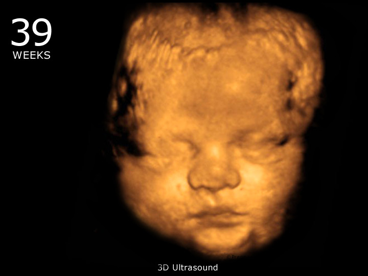
From 30 weeks the features that can usually be seen in this ultrasound include: your baby opening and closing their eyes, the amount of hair on their head (or lack thereof), their distinct facial features and whether they have mum or dad's nose! When can I do this ultrasound? between 26-40 weeks gestation.
Pregnancy due date tracker list, 13 weeks pregnant 3d ultrasound gender
Fetal ultrasound is used to check that the heart is working properly and to see if there could be any heart problems. The brain Below is an image of the base of the brain, called the cerebellum. This type of image usually is taken during an ultrasound done between 18 and 22 weeks of pregnancy.
The 26th week of pregnancy
At 26 weeks pregnant your body may be showing more evidence of all that growth and development in the form of stretching skin and possibly some new stretch marks. In today's post we are going to talk about your 26 week pregnancy and ultrasound.

O Baby! Murfreesboro, TN Gender Determination. 3D/4D Ultrasounds. 3d
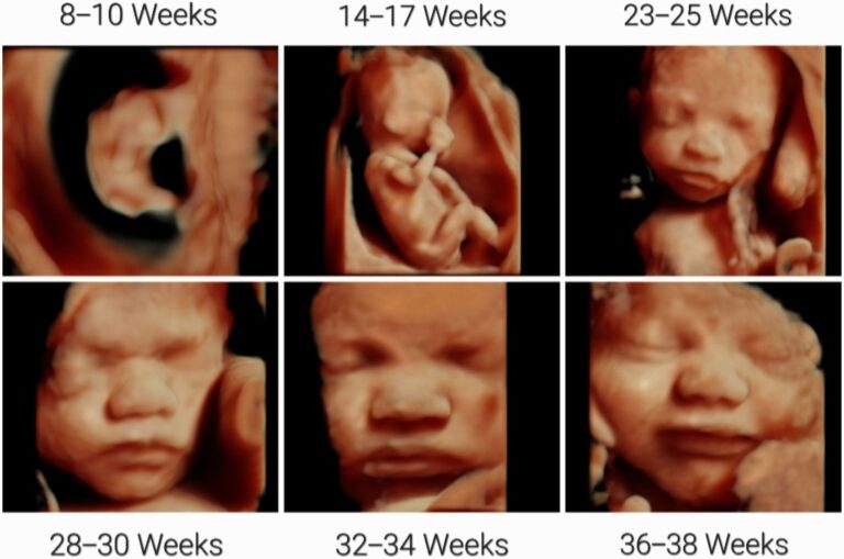
A 3D ultrasound involves taking thousands of slices in a rapidly occurring series called a volume of echoes. These send sound waves back at different angles, allowing for the characteristic 3D depth.. Generally, the recommendation is between 26 and 30 weeks gestation, unless otherwise suggested by your maternity care provider. By this time.

4d baby ultrasound miami Blimp Microblog Custom Image Library
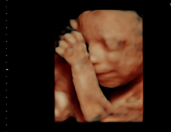
A 3D ultrasound makes it easier for doctors to interpret scans of the fetal heart anatomy. Depending on the technology used, a 3D baby ultrasound can even explore how the heart correlates with the vessels and structures around it. • Neural tube defects. The neural tube eventually becomes a baby's brain and spinal cord.

26 Weeks Ultrasound
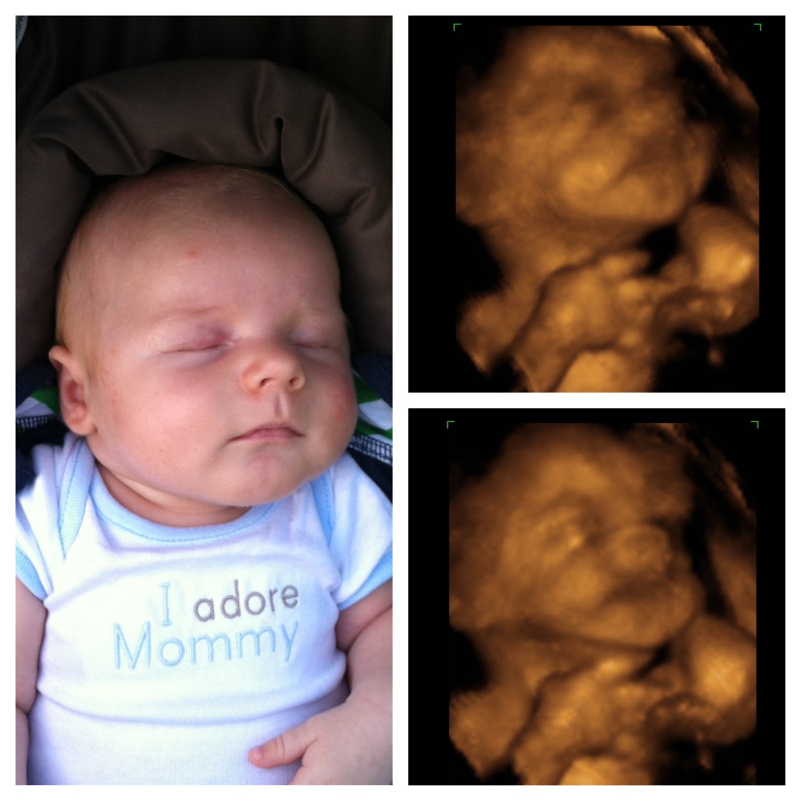
Here is a 3D Ultrasound of my baby girl at 26 weeks 5 days! Her eyes were open in the womb! I over her 3D ultrasound smile!Instagram:https://www.instagram.co.
26 Weeks Ultrasound
For bigger mothers, we suggest waiting until the baby reaches a certain size. We get better images after 26-27 weeks. Baby's movement could possibly decrease the quality of the 3D ultrasound pictures, especially if the movement is constant. But sometimes, the movements help us capture different poses or remove obscuring structures.
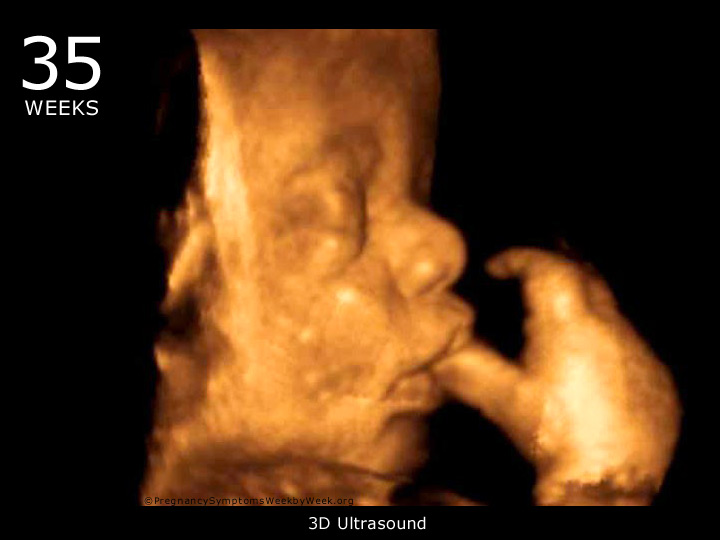
35 Weeks Pregnant ─ Pregnancy Symptoms Pregnancy Symptoms Week by
During pregnancy, regular ultrasounds are performed to monitor the development of the baby. One of the important ultrasounds is the 26 week ultrasound, which is usually done by the doctor. This ultrasound scan is typically done around the 26th week of pregnancy. It provides valuable information about the baby's growth and development.

When is the Best Time to Get a 3D Ultrasound? Mother Nurture Ultrasound
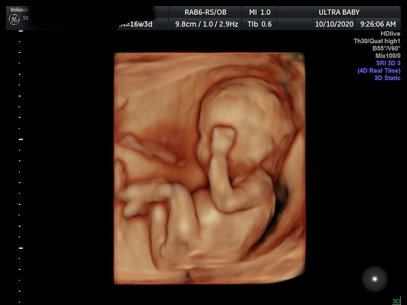
A video of our daughter when she was 26 weeks old while in utero. Taken by a 3d/4d Ultrasound machine.